Heart Chamber Size Echocardiography
Browse our collection of Heart Chamber Size Echocardiography templates. Each calendar is free to download and optimized for printing on standard paper sizes. Click any image to view the full-size version and download it instantly.
 Rotation Angle From 4 Chamber To 2 Chamber View In Echocardiography
  PDF Indium 111 Platelet Scintigraphy And Two dimensional
  Figure 1 From Echocardiographic Reference Ranges For Normal Cardiac
  Pin By Didi Ramos On Ultrasound Cardiac Sonography Diagnostic
  Patient s Echocardiography Showing Normal LV Size With Preserved
  CME Right Ventricle Right Atrium Size Echocardiography
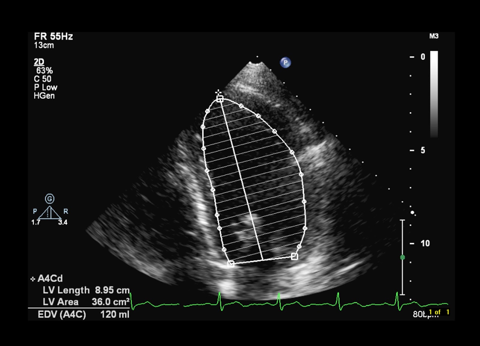 Lv Chamber Size Is Normal Keweenaw Bay Indian Community
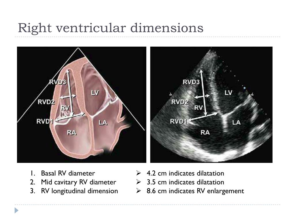 Normal Lv Dimensions Paul Smith
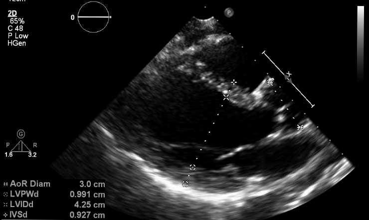 What Does An Echocardiogram Show MyHeart
 Pin On EcoMedCrit
 Echocardiogram echo Paula Moore M D Dysautonomia MVP Center
 2D Normal Values Of Cardiac Chambers And Inferior Vena Cava LVEDd
  Table 3 From Echocardiographic Reference Ranges For Normal Cardiac
  Apical 4 Chamber View Of A Normal Heart YouTube
 What Is An Echocardiogram Uses Procedure And Results
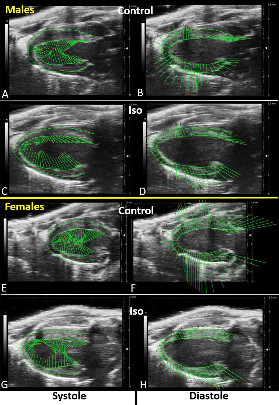 Probl m De Aspekt Echo Kg Ivs Neloj lnosti Uctievanie Minimalizova
  Standard Apical 4 Chamber 2D Transthoracic Echocardiography Images Of
  Table 4 From Echocardiographic Reference Ranges For Normal Cardiac
  Trans Esophageal Echocardiogram TEE
  Regional Myocardial Contractile Function Wall Motion Abnormalities
 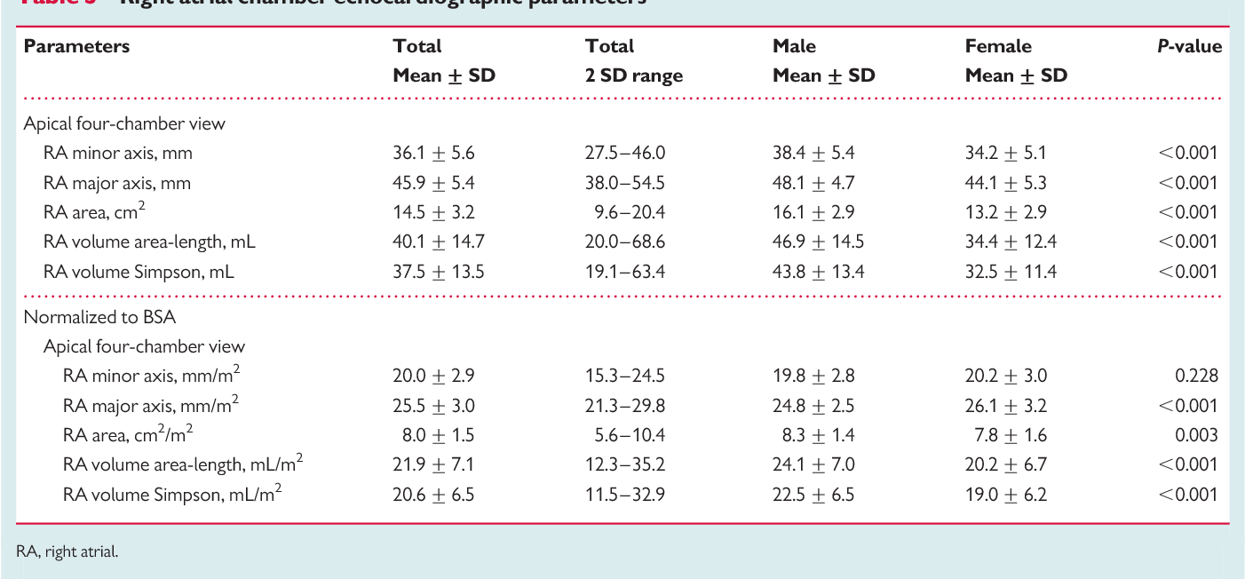 Table 2 From Echocardiographic Reference Ranges For Normal Cardiac
  Stem Cells In The Treatment Of Heart Failure MyHeart
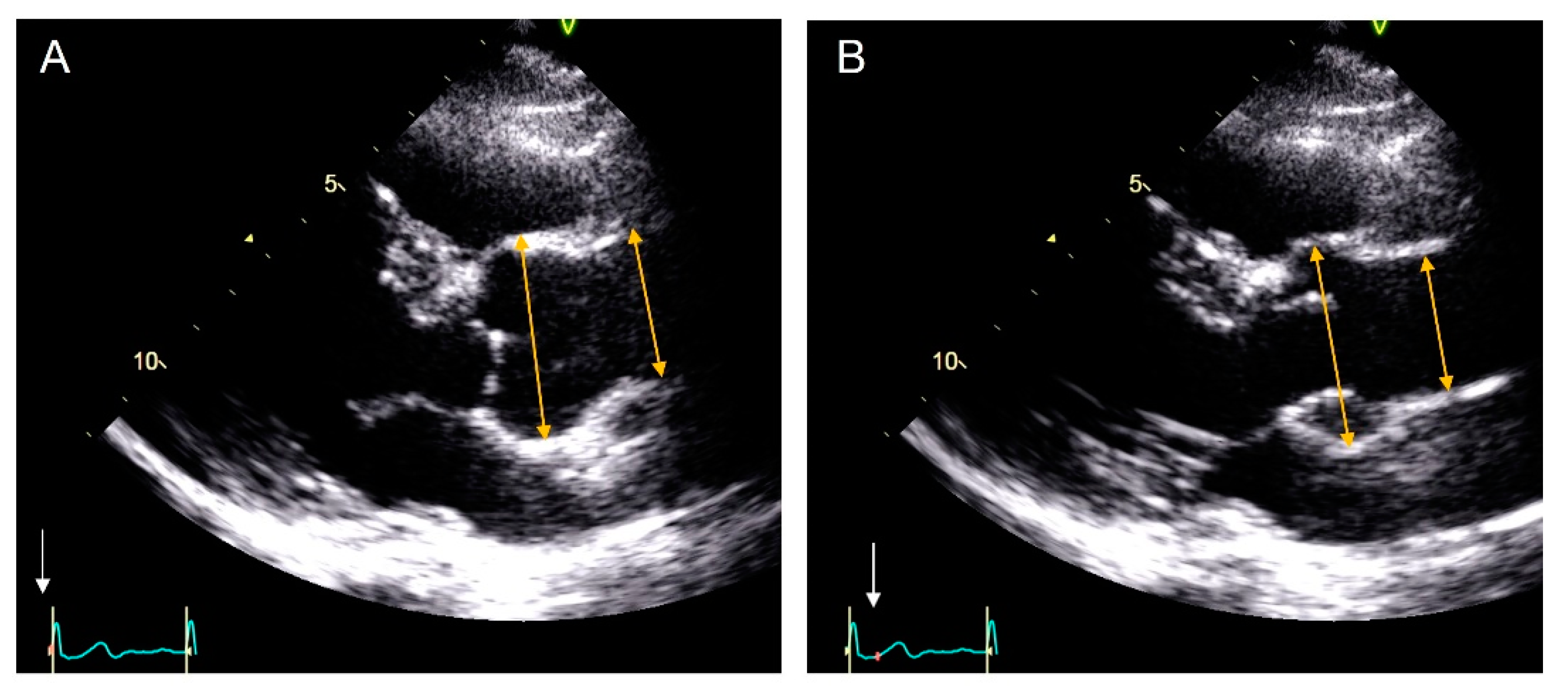 Krovia Dareb k Syndr m Measurement Of Aorta In Echo Atlantick
  Three Dimensional Echocardiographic Assessment Of Left Heart Chamber
  Ase Lv Chamber Size The Art Of Mike Mignola
 What Is The Normal RV Size How To Measure It By Echocardiography
  View Image
 Pocket size Echocardiography Device Effective For Thoracic Aortic
  Echocardiography Of The Ascending Aorta Steve Gallik
 Echocardiography Transesophageal Echocardiogram Ultrasonography
  Normal Values For The Right Ventricle RV Linear And Area Dimensions
  Locations Of Measurements By Echocardiography A ANN aortic Annulus
  Table 2 From Echocardiographic Reference Ranges For Normal Cardiac
 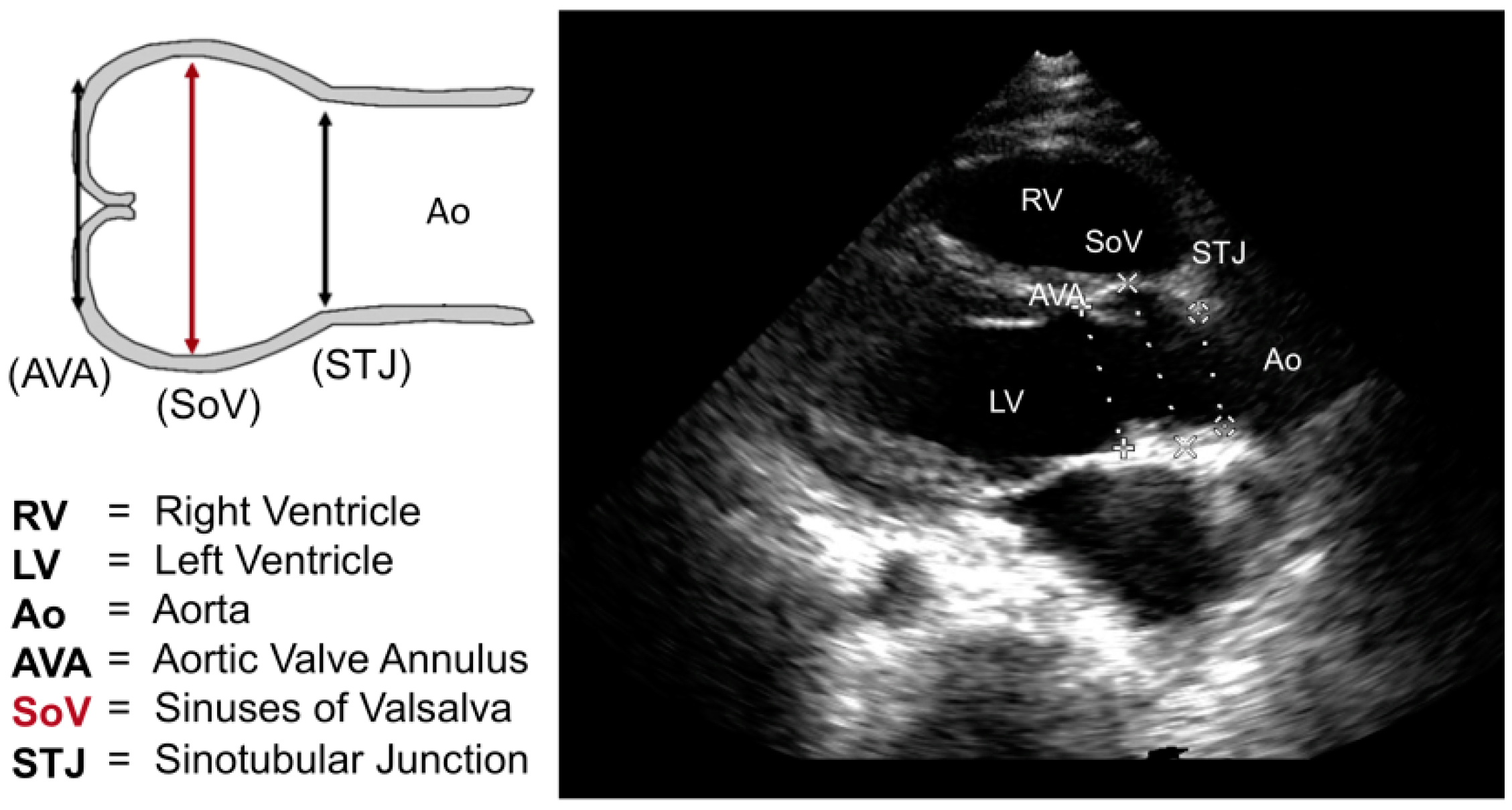 IJMS Free Full Text Aortic Root Dilatation In Mucopolysaccharidosis